New Park House Dental Centre has a commitment for being at the forefront of dental technology.
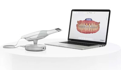
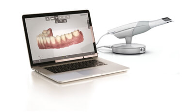
Intra-oral scanning has already revolutionised many aspects of dentistry. With the TRIOS
scanner we are not only able to scan the teeth and email the "impression" to our lab digitally but the digital data also links to many of the latest developments in implant planning, clear aligner orthodontics and smile design.
Other benefits include:
At New Park House Dental Centre our commitment to constantly develop our skills and use the latest technology has meant that a CT scanner is a must-have piece of equipment.
Having this advanced equipment available on site allows our patients to receive the most up to date dental care possible.
Our Cone Beam Computed Tomography scanner (CBCT) is a state-of-the-art scanning machine that produces highly accurate 3D images of a patient's mouth.
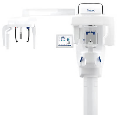
We consider CBCT scanning to be essential when planning many complex treatments such as implant placement and specialist endodontic treatments.
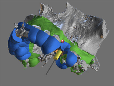
Combining the CBCT data with our Trios intra-oral scanner and the latest 3D printing technology allows us to create highly accurate surgical guides for our implant surgery. This can often result in more accurate placement and less invasive surgery.
All our dentists and many of our staff are trained to take high quality dental x-rays.
We audit the quality of our radiography on an annual basis to ensure we maintain the highest possible standards.
The high resolution digital images can be viewed almost instantly.
All surgeries have large wall mounted TV's to view the images and further help communication.
The use of these high resolution images allows us to:
Many patients have never seen their teeth in this level of detail and the feedback we always receive is that the increased understanding of their problems and the risks and benefits of the solutions, makes for a more relaxed treatment.
Most x-rays are taken to diagnose the cause of symptoms which cannot be seen by eye, and to monitor periodontal (gum) disease. The x-ray enables the dentist to give more accurate advice and discuss appropriate treatment options.
X-ray examinations are only performed when the risk of not having the examination is greater than the risk of having exposure to radiation.
Before having the x-ray your dentist will do a thorough clinical examination and the minimum number of radiographs (x-rays) will be taken.
We are all exposed to background radiation every day. It comes from our surroundings, such as rock, earth, building materials and the sun. Some radiation also comes from the food we eat.
A normal dental x-ray is equivalent to one day of average natural background radiation.
At New Park House we are subject to both national regulations and our own strict in-house procedures. The equipment we use is pre-set to the minimum level. All our equipment is maintained and are tested routinely for accuracy.
If you are concerned about having an x-ray please talk to your dentist so they can help.
Most dental x-ray films are angled away from the pelvic region and do not pose a risk.
That said, we normally avoid taking x-rays during pregnancy unless considered necessary by your dentist and with your permission. Your dentist will always discuss the risks and benefits with you.

All our dentists and many of our staff are highly trained in the use of digital SLR cameras, utilising a very specific setup for clinical dental photography.
The images taken allow our patients to see problems in the same detail as the dentist.
All surgeries have large wall mounted TV's to view the images and further help communication.
The use of these high resolution images allows us to:
Many patients have never seen their teeth in this level of detail and the feedback we always receive is that the increased understanding of their problems and the risks and benefits of the solutions, makes for a more relaxed treatment.
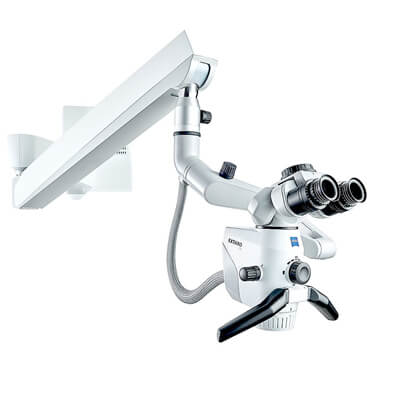
Since its inception in 1953, the surgical microscope has become firmly established in numerous medical fields across a broad range of clinical applications.
A large number of surgical procedures would now be totally inconceivable without the use of a microscope.
In dentistry, the microscope has also redefined the concept of visualisation. Particularly in the field of endodontics (root canal treatment), our specialist endodontist routinely uses our microscope.
| Monday | 8.30 - 17.00 |
| Tuesday | 8.30 - 17.00 |
| Wednesday | 8.30 - 17.00 |
| Thursday | 8.30 - 17.00 |
| Friday | 8.30 - 16.30 |